Seminar
A combined optical and atomic force microscope for materials and life sciences research
On July 23rd, 2024, Dr. Pham Thanh Tri visited the Research Center for Infectious Diseases and gave an insightful talk about how a combined optical and atomic force microscope can be applied in materials and life sciences research
Computer simulations and image analysis, light microscopy, atomic force microscopy, cell biophysics, biosensors, nanoparticles, bio/nanomaterials, and their applications in life sciences

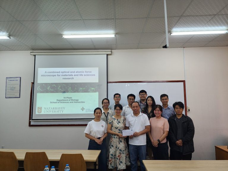
Seminar group photo
Have a look at the summary of Dr. Tri’s talk
In this talk, we demonstrated the capabilities of a hybrid optical-atomic force microscope, which has many applications in the fields of materials science and life sciences research. When operating in individual mode, time-lapse imaging, often referred to as live cell imaging, can be utilized to see where a specific organelle or protein of interest is located in a cell or tissue as well as its molecular activity. Membrane fibers and bacterial biofilms can also be observed under an optical microscope by labeling them with a fluorescent dye to determine their shape and size. In contrast to optical microscopy, atomic force microscopy was originally developed to image the morphology of a material surface, using a nano probe to physically scan the surface. Its current capacity, though, far exceeds what was originally intended. It can be used to measure the mechanical, electrical, and magnetic properties of the surface of both living and non-living samples in addition to mapping the surface mophorlogy. In the field of mechanobiology, it has also been utilized recently to measure the biophysical characteristics of cells and tissues in liquid. In a hybrid mode, an atomic force microscope can examine surface characteristics like morphology, adhesion, and stiffness, while an optical microscope provides a clear visualization of the area we are measuring and can reveal the localization or activity of a particular protein of interest through fluorescence emission. We can therefore correlate the molecular activity of the living samples with their biophysical characteristics thanks to this combined setup. During my presentation, I will provide several examples of how this hybrid setup is used extensively in materials and biological sciences research. The study areas include biosensors, gas sensors, photovoltaic solar cells, live cell imaging, bacterial biofilms, mechanobiology, nano/biomaterials, metal alloys, and their interactions with living cells.
